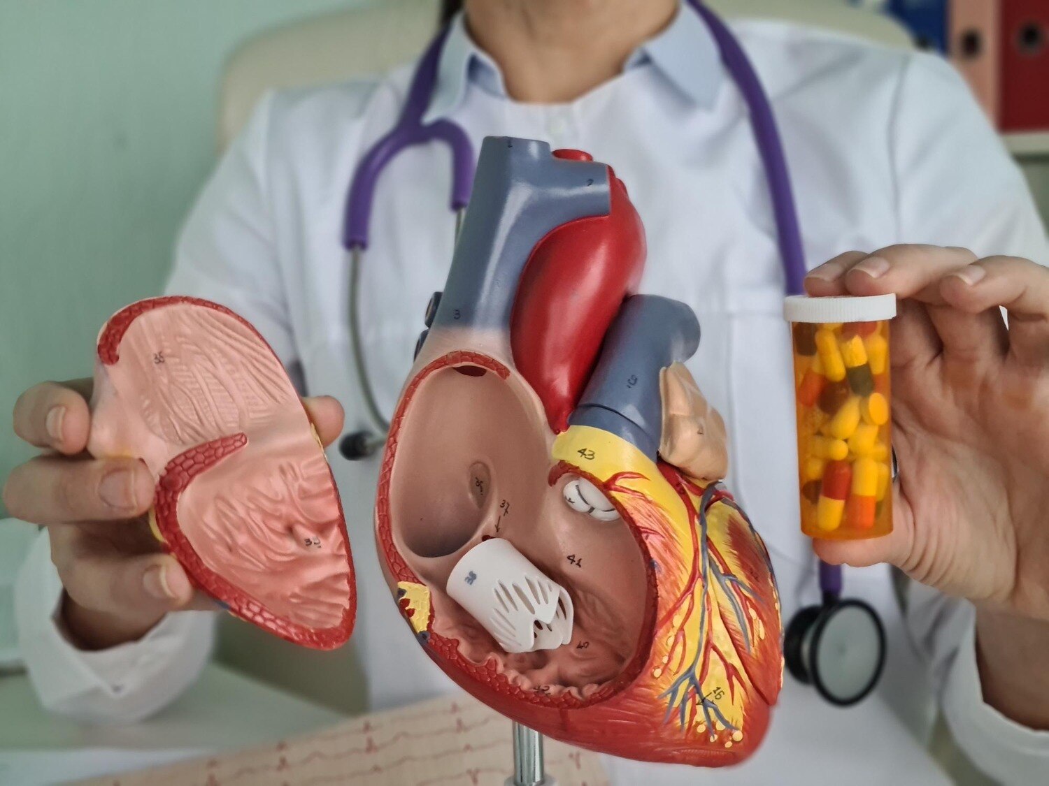
Treatment depends on which valve is affected, whether it is narrowed or leaking, how severely the heart is responding, and whether symptoms are present. By following the diagnostic pathway and comparing monitoring, medicines, repair, replacement, and catheter-based procedures, you can prepare for a more informed discussion with a cardiac team.
Key takeaways
- Breathlessness, chest pain, fainting, swelling, or palpitations need valve assessment.
- Echocardiography measures valve narrowing, leakage, chamber changes, and pressure effects.
- Treatment ranges from monitoring and medicines to repair, replacement, or catheter procedures.
- Ask for a coordinated plan covering diagnosis, intervention, anticoagulation, rehabilitation, and follow-up.
Which valve problem could be causing your symptoms?
A valve can become narrowed (stenosis), fail to close (regurgitation or insufficiency), bulge backward (prolapse), or fail to form or open (atresia). Causes include congenital defects such as bicuspid aortic valve, age-related calcium buildup, rheumatic heart disease, and infective endocarditis.
Secondary mitral regurgitation can result from heart failure, ischemia, or rhythm disease rather than primary valve damage.
| Valve | Main problems and common causes | Symptoms and clues |
|---|---|---|
| Aortic | Stenosis from calcium or bicuspid anatomy; regurgitation causes leakage from the aorta | Breathlessness with exertion, chest pain, dizziness, or fainting |
| Mitral | Prolapse or degeneration can cause regurgitation; rheumatic disease can cause stenosis | Breathlessness when lying down, fatigue, or palpitations |
| Tricuspid | Regurgitation often follows pulmonary hypertension, left-sided disease, right-ventricular enlargement, or atrial fibrillation | Ankle swelling, abdominal fullness, or unexplained weight gain |
| Pulmonary | Congenital stenosis or atresia; regurgitation can follow previous repair | Reduced exercise tolerance, breathlessness, or fatigue |
- Arrange assessment for a new murmur, persistent breathlessness, reduced exercise tolerance, palpitations, swelling, or unexplained weight gain.
- Seek emergency care for severe breathlessness at rest, chest pain, fainting, blue lips, or sudden worsening.
Echocardiography—not the murmur alone—shows valve structure, narrowing, leakage, and heart function, guiding valvular heart disease care.
How do doctors confirm valve disease and measure its severity?
A murmur is a sound, not a severity score. A valve disease doctor in Navi Mumbai will examine your pulse, blood pressure, heart sounds, breathing, neck veins, and ankle swelling, then review symptoms and exercise tolerance.
The examination can suggest stenosis or regurgitation, but it cannot show whether the valve is damaged or how hard the heart is working.
A transthoracic echocardiogram (TTE) usually confirms the lesion and measures valve opening, pressure gradients, regurgitant volume, ventricular size and ejection fraction, pulmonary pressure, and associated valve problems. A low gradient does not automatically mean mild aortic stenosis; low-flow, low-gradient disease can occur with reduced or preserved ejection fraction.
Further tests answer specific unanswered questions:
- ECG detects rhythm problems, prior heart strain, or conduction abnormalities; it does not measure valve narrowing.
- Transesophageal echocardiography gives clearer views when chest images are limited or before repair, replacement, or TEER.
- Cardiac CT measures anatomy and aortic-valve calcium; cardiac MRI measures chambers and regurgitation.
- Exercise testing or dobutamine stress echocardiography clarifies symptoms and selected low-flow, low-gradient aortic stenosis.
- Cardiac catheterisation measures pressures directly and helps plan intervention when non-invasive results conflict.
New breathlessness, fainting, chest pain, declining exercise tolerance, or swelling warrants reassessment rather than waiting for the next routine scan. Severity and ventricular response determine surveillance intervals.
Which treatments fit aortic, mitral, tricuspid, and pulmonary valve disease?
Treatment follows the valve lesion, its severity, symptoms, ventricular effects, and anatomy. Mild disease with stable heart function may need surveillance with repeat echocardiography, while severe symptomatic disease often needs an intervention. Medicines relieve congestion, control blood pressure or rhythm, but they do not open a severely narrowed valve or permanently stop a major leak.
| Valve | Common treatment choices | When they fit |
|---|---|---|
| Aortic | Medicines; SAVR; TAVR; balloon valvotomy | Medicines control symptoms or blood pressure. SAVR or TAVR treats severe symptomatic aortic stenosis; SAVR is standard for many cases of severe aortic regurgitation. Balloon treatment is usually a temporary bridge or special-case option. |
| Mitral | Surveillance; diuretics and heart-failure treatment; repair or replacement; TEER; balloon commissurotomy | Repair is preferred for suitable primary degenerative regurgitation. TEER suits selected severe symptomatic patients at high surgical risk. Balloon commissurotomy treats suitable rheumatic mitral stenosis. |
| Tricuspid | Diuretics; treatment of rhythm, lung or left-heart disease; repair or replacement | Medicines reduce swelling while the cause is addressed. Severe symptomatic disease may require surgical or transcatheter repair or replacement. |
| Pulmonary | Surveillance; balloon valvotomy; repair or replacement | Balloon valvotomy commonly treats suitable congenital pulmonary stenosis. Severe regurgitation or complex congenital disease may require repair or replacement. |
Infective endocarditis needs urgent blood cultures, intravenous antibiotics and assessment for surgery when infection damages the valve, causes heart failure or persists.
Valve disease treatment options in Navi Mumbai should therefore include specialist echocardiography, rhythm and heart-failure assessment, CT or catheterisation when needed, and referral to a multidisciplinary valve team for TAVR, TEER, repair or replacement.
How are valve repair, replacement, anticoagulation, and follow-up decisions made?
Severe symptomatic aortic stenosis needs valve replacement; medicines alone cannot remove the obstruction. The choice between catheter treatment, surgery, repair, or replacement depends on anatomy, age, life expectancy, surgical risk, valve durability, pregnancy plans, bleeding risk, and your informed preference.
| Option | Main trade-off | When it applies |
|---|---|---|
| SAVR | Open surgery with established durability | Younger or low-risk patients, bicuspid valves, aorta disease, or anatomy unsuitable for TAVR |
| TAVR | Catheter-based treatment with faster recovery, but different durability and pacemaker considerations | Selected aortic stenosis patients after CT-based anatomical and access assessment |
| Mitral repair | Preserves the native valve and usually avoids prosthetic-valve complications | Primary degenerative mitral regurgitation when an experienced team expects a durable repair |
| Mitral replacement | Reliable correction when repair is unlikely to last | Heavily damaged valves, failed repair, or unsuitable anatomy; TEER is reserved for selected high-risk symptomatic patients |
Mechanical valves last longer but require lifelong warfarin and INR monitoring, with clotting and bleeding risks. Bioprosthetic valves usually avoid lifelong anticoagulation solely for the valve but can wear out; age, future valve-in-valve treatment, pregnancy plans, and bleeding risk affect the decision.
- Use a vitamin-K antagonist such as warfarin for a mechanical valve or atrial fibrillation with rheumatic mitral stenosis; do not substitute a direct oral anticoagulant without specialist advice.
- Repeat echocardiography at the interval set by valve severity, and report breathlessness, fainting, swelling, fever, or reduced exercise promptly.
- Maintain dental hygiene, treat infections early, and ask whether infective-endocarditis antibiotics are indicated before a dental procedure.
- A valve specialist in Navi Mumbai should coordinate echocardiography, ECG, rhythm and heart-failure assessment, CT or catheterisation, and referral to a multidisciplinary valve team when intervention is needed.
What should a complete valve-care pathway in Navi Mumbai include?
A complete local pathway should connect diagnosis to a treatment decision, not stop at a murmur or one scan. For heart valve treatment Belapur, check that the service can arrange specialist transthoracic echocardiography, ECG, rhythm and heart-failure assessment, then escalate imaging when anatomy or symptoms remain unclear.
Severe cases need intervention planning and referral to a multidisciplinary valve team.
| Pathway checkpoint | What to verify | Why it matters |
|---|---|---|
| Specialist echocardiography | Valve gradients, regurgitant volume, chamber size, ventricular function and pulmonary pressure | Confirms severity rather than relying on the murmur |
| Rhythm and heart-failure review | ECG, rhythm monitoring when indicated, fluid assessment and medication review | Finds atrial fibrillation or ventricular strain that changes treatment |
| Advanced imaging | Transesophageal echo, CT, cardiac MRI, stress testing or catheterization when needed | Clarifies anatomy, symptoms and procedural risk |
| Intervention planning | Repair, replacement, TAVR or TEER options reviewed against anatomy, age, risk and preference | Prevents a procedure chosen without complete assessment |
| Multidisciplinary referrals | Cardiologist, cardiac surgeon, imaging specialist and heart-failure or rhythm specialist | Brings the right expertise together for complex decisions |
- Who interprets the echocardiogram, and can I receive the measurements?
- Why do I need another scan or catheterization?
- Is my valve problem primary, or is heart failure or rhythm disease driving it?
- Which team will review repair, replacement, TAVR or TEER?
- What happens if symptoms continue after treatment?
When comparing Heart Fitt Cardiac Wellness Clinic with another local option, ask who coordinates these steps and where procedures are performed.
Related service
Valve Disease Treatment Dr. Charan Reddy at Heart Fitt Cardiac Wellness Clinic, Belapur, specializes in comprehensive valve disease treatment. View service → |
Frequently asked questions
Which valve problem could be causing my symptoms?
Stenosis restricts blood flow, while regurgitation lets blood leak backward. Aortic, mitral, tricuspid, and pulmonary valve disease can cause breathlessness, fatigue, chest discomfort, fainting, swelling, or palpitations. Congenital defects, calcium buildup, rheumatic disease, prolapse, and infection are possible causes.
How do doctors confirm valve disease and measure its severity?
Doctors combine your history and examination with tests such as an ECG, chest X-ray, transthoracic echocardiogram, Doppler measurements, and sometimes transesophageal echocardiography, stress testing, cardiac CT, cardiac MRI, or cardiac catheterisation. These tests assess valve opening, leakage, blood-flow speed, chamber size, pumping function, and pressure effects.
Which treatments fit aortic, mitral, tricuspid, and pulmonary valve disease?
Treatment depends on the affected valve, whether it is narrowed or leaking, symptom burden, ventricular changes, anatomy, age, surgical risk, and cause. Options include monitoring, medicines for congestion or rhythm problems, balloon procedures, surgical repair, surgical replacement, and selected transcatheter interventions.
How are valve repair, replacement, anticoagulation, and follow-up decisions made?
Repair is preferred when it can provide a durable result, especially for suitable mitral or tricuspid disease. Replacement may use a mechanical or tissue valve, each with different durability and anticoagulation implications. Follow-up includes repeat echocardiograms, symptom review, medication checks, dental and infection advice, and urgent assessment for worsening symptoms.
What should a complete valve-care pathway in Navi Mumbai include?
Look for coordinated evaluation by a valve disease doctor in Navi Mumbai, access to echocardiography and other imaging, clear severity grading, referral for intervention when needed, medication and anticoagulation management, rehabilitation guidance, and scheduled follow-up near Belapur or your home.
Keywords